
A microscope and a telescope combine an objective with an eyepiece, but their input geometry differs. The microscope examines a nearby object; the telescope typically receives nearly parallel light from a distant scene. Following the intermediate image explains why their magnification formulas differ and which distances must be controlled.
A microscope’s intermediate image
Consider two positive thin lenses in air, separated by . In a finite-conjugate microscope, the objective forms a real intermediate image. For relaxed viewing, that image sits at the eyepiece’s front focal plane, so light leaves the eyepiece parallel. If their focal lengths are and :
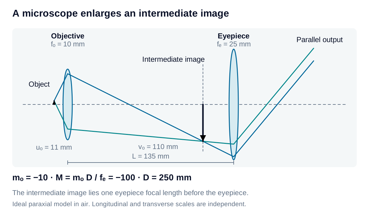
The eyepiece acts as a magnifier, with relaxed-view angular gain relative to viewing the intermediate image unaided at distance . Comparing the complete instrument with the original specimen viewed unaided at gives:
Unlike , which compares physical image heights, compares apparent angles. The conventional reference mm is a comparison distance, not an assumed capability of every eye. All distances must use the same units.
Derivation: microscope magnification combines image scale and viewing angle
Let the specimen height be and the intermediate-image height be . The objective’s finite image has transverse magnification . For relaxed viewing, the intermediate image is one eyepiece focal length in front of the eyepiece, so the lens separation satisfies:
Applying the objective’s thin-lens equation gives:
Use consistent signed apparent angles for the specimen and its viewed image. In the paraxial approximation, the eyepiece presents an apparent angle . The unaided specimen at reference distance subtends . Thus:
The objective contributes a physical image-height ratio; the eyepiece contributes the angular factor . Multiplying two finite-image transverse magnifications would describe a different output condition. A camera placed at the intermediate image records the objective’s image scale and does not automatically acquire the eyepiece’s visual angular factor.
For mm, mm and mm, the objective image is at 110 mm and the object is at 11 mm. Thus and : the view is inverted. A real intermediate image requires ; enlargement requires . Infinity-corrected microscopes use a tube lens and need their own optical model.
An afocal telescope
A Keplerian telescope uses two positive lenses. For a distant scene and relaxed viewing, the objective’s rear focal plane coincides with the eyepiece’s front focal plane:
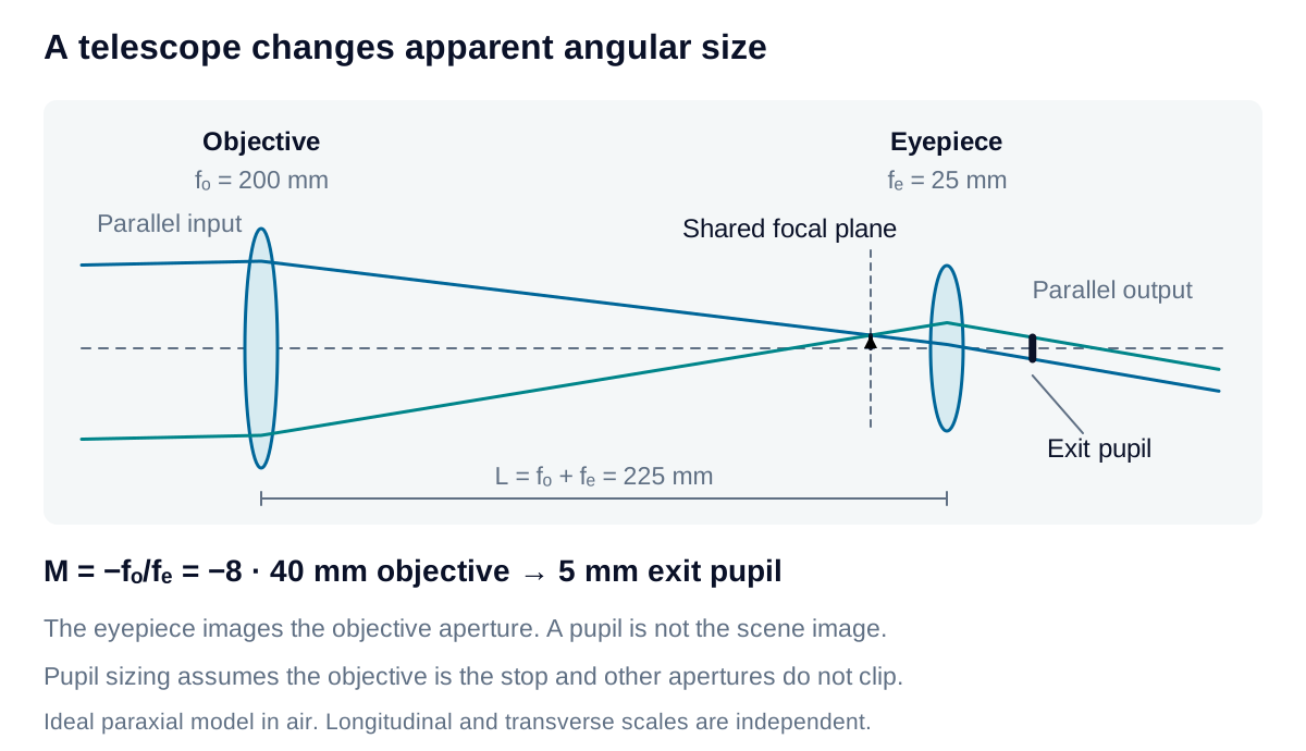
For a distant object with small full angular extent in radians, its intermediate-image size is:
That real image lies at the objective’s rear focal plane. The infinity-input model specifies angular extent rather than a finite object height and distance, so an ordinary objective transverse magnification is not reported.
Derivation: telescope angular magnification and intermediate-image size
A distant object supplies a parallel ray bundle for each field point. Let be the signed paraxial propagation angle of one bundle. The objective forms its image at height in the rear focal plane. With that plane at the eyepiece’s front focus, the outgoing angle is:
Expressing both angles as viewing directions reverses both signs, leaving their ratio unchanged. Hence visual angular magnification is . The negative sign denotes inversion.
A centered object spanning full angle has edges at . Their image-height separation is approximately:
Here is a positive full size, not a signed image height. Convert degrees to radians before calculating: . This is a paraxial field estimate, not a wide-angle distortion model. A final afocal output has an angular image; the intermediate real image still has a definite physical size.
Exit pupil size and position
The exit pupil is the eyepiece’s image of the aperture stop. If the objective aperture is the stop and downstream optics do not clip the rays, its diameter gives:
Both quantities here are aperture diameters, distinct from the microscope’s reference viewing distance. Pupil size describes light delivery to the observer rather than scene-image size or resolution.
Derivation: the eyepiece images the objective aperture into the exit pupil
For pupil imaging, the objective aperture is the eyepiece’s object. Its distance from the eyepiece is . Apply the thin-lens equation to this aperture image:
This positive distance locates the ideal exit-pupil plane downstream from the eyepiece. The signed transverse magnification of the pupil image is:
A diameter is positive, so . Combining this with gives the reciprocal diameter ratio. This is magnification of the aperture image, separate from magnification of the scene. Real eyepiece assemblies require their actual pupil and principal-plane data to establish usable eye position.
With mm and mm, the separation is 225 mm and . An object spanning forms an intermediate image approximately 1.75 mm across. A 40 mm objective gives a 5 mm exit pupil, located 28.125 mm beyond the ideal eyepiece plane under the stated stop assumption.
Greater apparent size does not guarantee additional detail. Objective aperture, wavelength, aberrations, field coverage, and pupil access still constrain useful performance. For camera-based measurement, specify the sensor-plane image scale separately from the visual angular magnification quoted for an eyepiece.